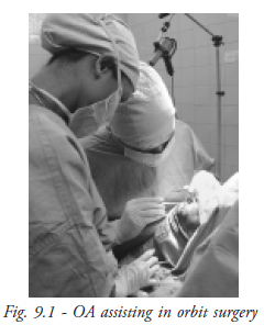
Role of OA in assisting speciality surgeries
Orbit - sac surgeries
DCR
Pre op work up
- Syringing / Probing
- BP
- RBSL
- Blood tests like : Hb, BT,CT, Blood group and type, ESR
- HIV
- Counselling / consent
Steps involved in the surgery
- Cleaning : The eye and the nose and most of the cheek and forehead are cleaned
- Draping : Two sterile towels are used for draping,one partially wrapped around the head and covering the other eye, and the other covering the nose and mouth up to the chest. Towel clips/artery forceps are used for anchoring the drapes
- Nasal cavity is packed with adrenaline soaked guaze after putting xylocaine jelly
- Skin incision given
- Blunt dissection is done
- The Medical canthal xendon is divided
- Periosteum is reflected off the nasal bone
- Osteotomy is made and enlarged
- Probe is passed to locate the lacrimal sac
- The anterior sac mucosa is cut to form a rectangular flap. The posterior part of the sac is excised
- The nasal mucosal flap is made
- The two flaps are anastomosed using three interrupted sutures of 6-0 vicryl
- Duct patency is checked
- Muscle layer is closed with 6-0 vicryl and skin with 4-0 silk
Instruments
- For nasal packing
- - Thudicums nasal speculum
- - Nose packing forceps
- - Nose pack (Thin length of gauze soaked in Adrenaline 1:100,000)]
- For local infiltration
- - A 5 cc syringe with disposable needle (with 3-5 cc of 2% xylocaine)
- For surgery
- - No 15 blade with BP handle
- - Artery forceps
- - Cat's paw retractors
- - Freer periosteal elevator
- - Forceps – toothed, plain
- - Kerrison Bone punch
- - Punctum dilator
- - Probe
- - No 11 blade on BP handle
- - Westcott spring handled scissors
- - Needle holder
- - 6-0 vicryl and 4-0 silk sutures on needle
- - 2cc syringe with cannula for syringing
Dacryocystectomy (DCT)
Steps
- Painting and draping is done
- Local anaesthetic is injected
- Skin incision is put in medial canthal area with no.15 blade
- Wound retracted with cat's paw retractors
- Blunt dissection is done with mosquito and artery forceps and lacrimal sac is identified after probing
- The sac is separated from surrounding tissue with sharp and blunt dissection and removed.
- Betadine wash is given
- The wound is sutured with 4-0 silk
Instruments
- No.15 blade
- Two cat's paw retractors
- Mosquito forceps
- Sac dissector
- Toothed forceps
- Bowman's probe
- Scissors
- Artery forceps
- 4-0 silk suture
Special considerations
- Pre operative
- As DCR (like most orbital and lacrimal surgeries) may involve significant blood loss, pre-op testing (like BP, Hb, BT, CT, HIV, Blood grouping and typing) is very important.
- Conditions like bleeding disorders, uncontrolled diabetes and hypertension, cardiac disease,asthama etc should be properly treated before the patient is taken up for surgery. If such conditions are present a physician's opinion should be taken and fitness for surgery confirmed.
- The surgery may be done under general anaesthesia or local anaesthesia. For children,mentally unfit or deaf patients, general anaesthesia is given. For apprehensive patients or people with low pain tolerance, counselling should be done. If reasonably co-operative, local anaesthesia or IV sedation are used, otherwise general anaesthesia is planned.
- Intra operative
- The surgical field has to be kept as clear as possible. Oozing blood has to be continuously mopped. Suction or cautery may have to be used and should be kept ready.
- The patient should be monitored throughout the surgery. If available, pulse oximeter should be attached before local anaesthesia is given.
- In case of severe Intra operative bleed, the patients' BP should bes checked and controlled. BP apparatus and O2 cylinder should be available in Operation theatre.
- Intubation should be available as it may be required in case of intra-operative complications like loss of flaps etc.
- Post operative
- Some patients may develop bleeding through nose/mouth in the post-op period. If the blood is mostly clotted, the patient should be reassured and instructed to lie supine for a while. BP should be checked and controlled.
Glaucoma surgeries
Trabeculectomy
Surgical procedure- Conjuctival flap is made, fornix/ limbal based
- Cautery applied
- Triangular flap 4mm 3mm made
- Antimetabolites MMC_0.20% 5FU/5. ML. if indicated, are applied and washed with ringer Lactate
- Scleral flap is dissected out carefully up to the base
- Paracentesis done
- A/C ( Anterior chamber ) entered
- The trabecular meshwork is punched with Kelly's punch
- Peripheral Iridectomy (PI) done
- A/C formed
- Flap sutured with 10 ‘o' nylon suture
- Conjunctival flap sutured with 8 ‘o' vicryl
- S/C inj. Garamycin ½ cc+ inj, Decadron ½ cc given
Trabeculectomy surgery
Instruments- Speculum: Lid speculum is used to separate the lids as gently as possible without causing distress to the patient, pressure on the globe or interference with the surgery
- Superior rectus needle holder: It is used to take superior rectus bridle suture during surgery
- Toothed forceps
- Superior rectus forceps
- Plain on tying forceps
- Artery clamp: To secure the superior rectus bridle suture to the drape
- Conjunctival scissor: Used for making conjunctival flap either fornix based/ limbal based during surgery
- Corneal forceps: For holding delicate tissue like cornea during suturing
- No.15 blade on BP handle
- Blade breaker with razor blade
- Kelly's punch
- Iris holding forceps
- Vannas scissors
- Cautery: Used to cauterize bleeding vessels after making conjunctival flap under Saline (wet cautery )
- Swab stick: Used to clean the after surgery
- Mitomycin: 2mg/vial 0.2%. Taken on small sponge.
Trabeculectomy with ECCE
Surgical procedure- Conjunctival flap is made
- Cautery applied
- Triangular flap 4mm 3mm made
- Antimetabolites MMC_0.20% 5FU/5. ML. if indicated, are applied and washed with ringer Lactate
- Scleral flap is dissected out carefully up to the base
- The groove is extended on both sides along the limbus
- The AC is entered just beneath the flap
- Visco is injected
- Capsulotomy is made
- The section is extended (6 to 6.5 mm)
- The nucleus is delivered by pressure – counter pressure technique
- The cortex is washed
- The IOL is inserted
- PI is done
- Wound closed with 10-0 nylon
- Conjunctiva closed with 8-0 vicryl
Instruments
Same as for trab and including the instruments for ECCE
Cornea surgeries
Penetrating keratoplasty
- The donor eye is collected from the eye bank by the OA.
- The OA checks the case sheet and the eye to be operated on.
- Retro bulbar block (or GA ) is administered. The eye should not be massaged.
- Cleaning and draping of the part is done.
- Lid speculum is placed.
- Superior rectus bridle suture is put.
- The part to be excised is marked on the recipient cornea using a trephine ( usually 7 to 7.5mm ).
- The donor corneal button is excised using a larger trephine. The donor corneal button should be 0.5mm larger than the corneal button removed from the recipient eye.
- The damaged cornea (previously marked on the recipient eye ) is removed using corneal scissors and fine toothed forceps.
- The anterior chamber is formed by visco.
- The donor corneal button is sutured to the recipient eye using 10-0 nylon .
- The anterior chamber is washed and reformed with saline.
- The eye is patched after putting appropriate drops.
Instruments
- Lid speculum
- Superior rectus forceps with needle and thread
- Toothed forceps
- Plain forceps
- Trephine (2 are required, 1 is 0.5 mm larger)
- Corneal scissors
- 10-0 nylon suture
Pterygium excision with autograft
Steps of surgery- Cleaning and draping
- Lid speculum
- Stay suture (4-0 or 6-0 silk)
- Head and neck of pterygium is excised using a no.15 blade
- The appropriate size of the graft is marked on the conjunctiva in the supero temporal quadrant
- Saline is injected under the conjunctiva in the marked area
- The conjunctiva is lifted up and excised in the marked area.
- The graft is placed over the bare sclera and sutured with 6-0 vicryl
- The stay suture is removed
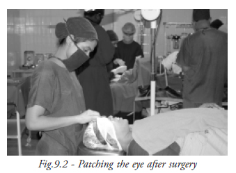
- Appropriate drops are applied and the eye is patched (Fig. 9.2).
- Lid speculum
- Plain forceps
- Toothed forceps
- No.15 blade
- BP handle
- Marker pen
- Conjunctival scissors
- 6-0 vicryl suture
The OA learned about the instruments to be used in the different speciality surgeries and the steps involved in each type of surgery.
Student exerciseAnswer the following
- What are the steps involved in the DCR surgery?
- List the instruments needed for nasal packing.
- List the instruments needed for trabeculatomy surgery.
- Write a short note on penetrating keratoplasty.
- What are the steps involved in the Pterygium excision with autograft?